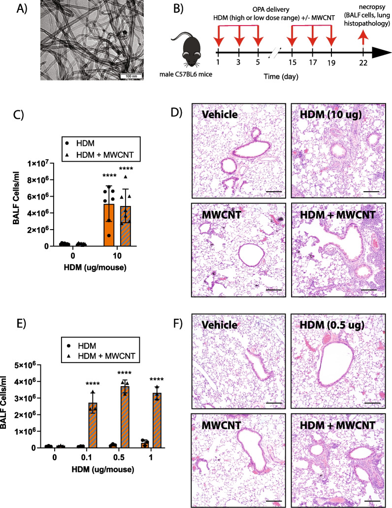
Fig. 1.
Comparison of high- and low-dose HDM extract on MWCNT-induced lung inflammation in male wildtype (WT) C57BL/6J mice. A TEM image of NC7000 MWCNTs used in this study. B Graphical representation of the experimental protocol used for Experiment 1 using a high dose of HDM extract (panels C and D) and experiment 2 using a low dose range of HDM extract (panels E and F). C Total inflammatory cell numbers in the BALF collected from mice exposed to high dose of HDM extract (10 µg per dose equivalent to 0.4 mg/kg per dose) with or without MWCNT (12.5 µg per dose equivalent to 0.5 mg/kg per dose) via oropharyngeal aspiration. n = 7. ****p < 0.0001 when comparing groups with and without MWCNTs as determined by two-way ANOVA with Tukey’s post hoc analysis. D Representative images of hematoxylin and eosin-stained lung tissue sections of mice treated with vehicle, high dose of HDM extract, MWCNTs, or combination of both. Black bars indicate 100 µm. E Total cell counts in BALF collected from mice exposed to low doses (0.1 µg, 0.5 µg, or 1 µg per dose) of HDM extract with or without MWCNTs (12.5 µg per dose) via oropharyngeal aspiration. n = 3. ****p < 0.0001 when comparing groups with and without MWCNTs as determined by two-way ANOVA with Tukey’s post hoc analysis. F Representative images of hematoxylin and eosin-stained lung tissue sections of mice treated with vehicle, low dose of HDM extract (0.5 µg per dose), MWCNTs, or combination of both. Black bars indicate 100 µm

