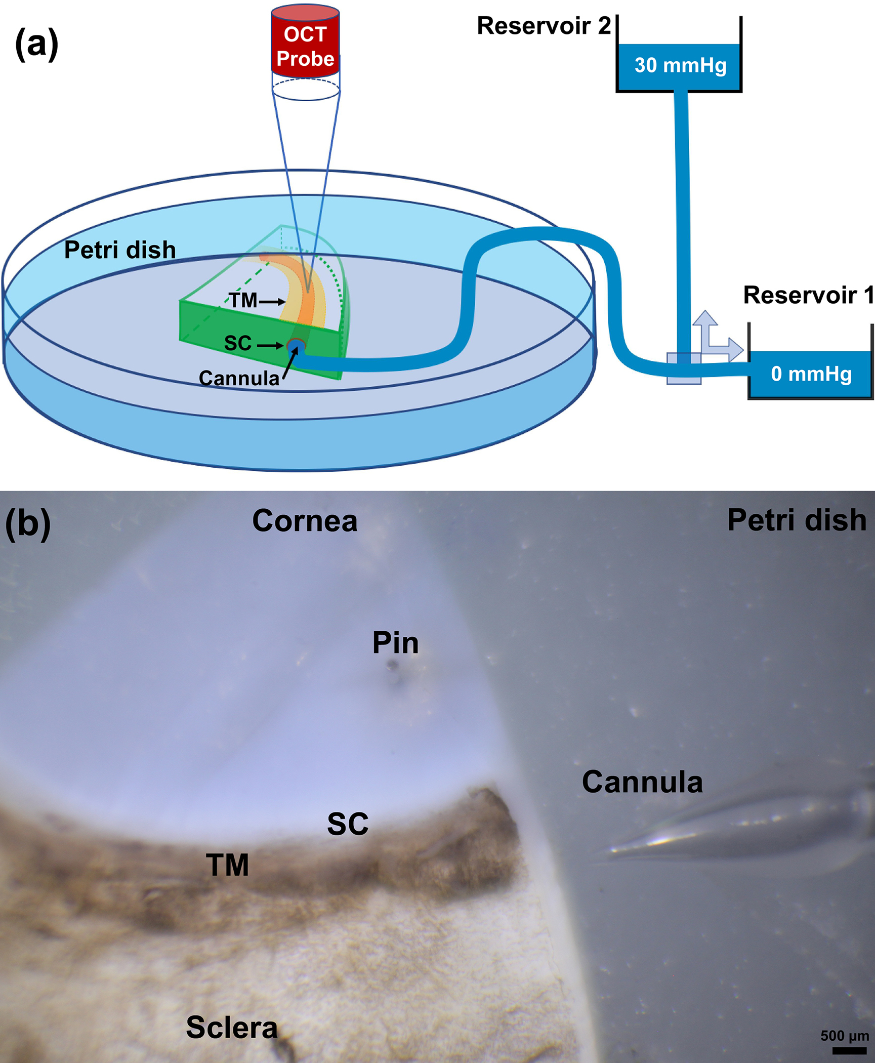
Fig. 1.

(a) Schematic overview of experimental setup, including the spectral domain optical coherence tomography (SD-OCT) system, two reservoirs used for controlling pressure in Schlemm’s canal (SC). Continuous acquisition SD-OCT B-scan images were acquired through the trabecular meshwork (TM), juxtacanalicular tissues (JCT) and SC at 30 Hz, resulting in a very high resolution images from which TM/JCT/SC motion can be determined in real time [132]. (b) A quadrant of the eye pinned in a petri dish and the structure of the cannula immediate before cannulation.
