Table 2.
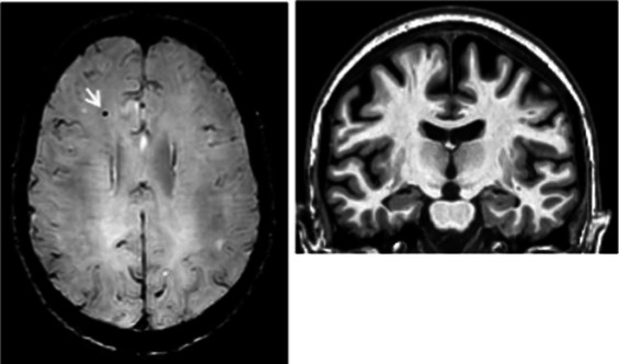
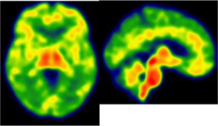
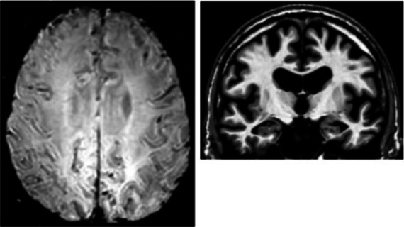
Dissociation between clinical and neuroinflammatory PET profiles in early AD.
| Clinical findings | CSF and APOE | SWI and T1-weighted MRI scans | Proposition of ongoing neuroinflammatory processes |
|---|---|---|---|
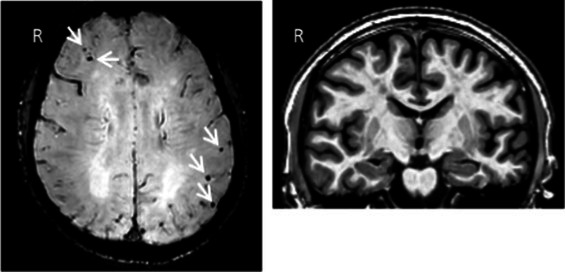
| Case 5: a 64 y.o. man who was referred for a memory complaint. At screening, he had 24/30 MMSE and impairment on episodic memory, denomination and categorical verbal fluency tests. On MRI, multiple lobar microbleeds without hemisiderosis were observed as well as WMH (Fazekas’s score of 8/9) and moderate cortical atrophy. | Aβ42: 208 P-tau: 184 T-tau: 1449 APOE E3/E3 TSPO MAB |
|
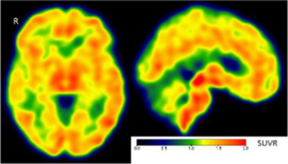 Toxic neuroinflammation associated with mixed angiopathy and AD pathological progression. |
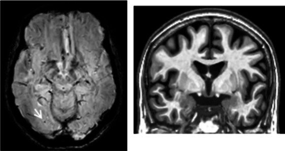
| Case 21: a 59 y.o. woman with early onset symptoms and familial history of AD. At screening, she had 23/30 MMSE and impairment on episodic memory, executive functions, processing speed and categorical verbal fluency tests. Three lobar microbleeds, WMH (Fazekas’s score of 3/9) and moderate cortical atrophy were observed on MRI. | Aβ42: 462 P-tau: 140 T-tau: 768 APOE E3/E4 TSPO MAB |
|
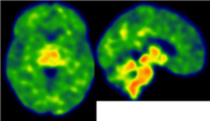 Low cortical neuroinflammation compared to the cerebellar cortex. |
| Case 2: a 66 y.o. woman with familial history of AD who was referred for a memory complaint. At screening, she had 30/30 MMSE, preserved memory, executive functions and processing speed but encoding impairment in visual recognition memory as well as decreased scores on long-term forgetting tests. Two lobar and one deep microbleed without hemosiderosis, WMH (Fazekas’s score of 5/9) and moderate cortical atrophy were observed on MRI. | Aβ42: 327 P-tau: 79 T-tau: 479 APOE E2/E4 TSPO HAB |
|
 Protective neuroinflammation that might be compensatory to the amyloid load in the frontal and cingulate regions in the absence of spread tau pathology and neurodegeneration. |
| Case 12: a 64 y.o. man with early onset atypical AD in a posterior cortical atrophy variant. He presented a familial history of AD. At screening, he had 21/30 MMS, multi-domain cognitive impairment, especially constructive apraxia and visual apperceptive agnosia. WMH (Fazekas’s score of 5/9) and cortical atrophy were observed on MRI. | Aβ42: 481 P-tau: 103 T-tau: 669 APOE E3/E3 TSPO HAB |
|
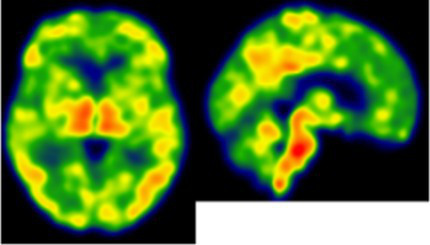 Toxic neuroinflammation associated with AD pathological progression, especially in posterior cortical regions. |
All fourth patients are right-handed. TSPO PET imaging showed SUVR relative to the cerebellar cortex and is represented in standard space in the same slice and intensity scale, whereas MRI scans are shown in native space. Cerebrospinal fluid AD biomarker values were abnormal for the four patients (see the method section for details).
Aβ42, amyloid-β 42; AD, Alzheimer’s disease; APOE, apolipoprotein E; CAA, cerebral amyloid angiopathy; CSF, cerebrospinal fluid; MMS, mini-mental state examination; MRI, magnetic resonance imaging, P-tau, phosphorylated tau; SWI, susceptibility-weighted imaging; T-tau, total-tau; TSPO, translocator protein; WB, whole brain; WMH, white matter hyperintensities.
