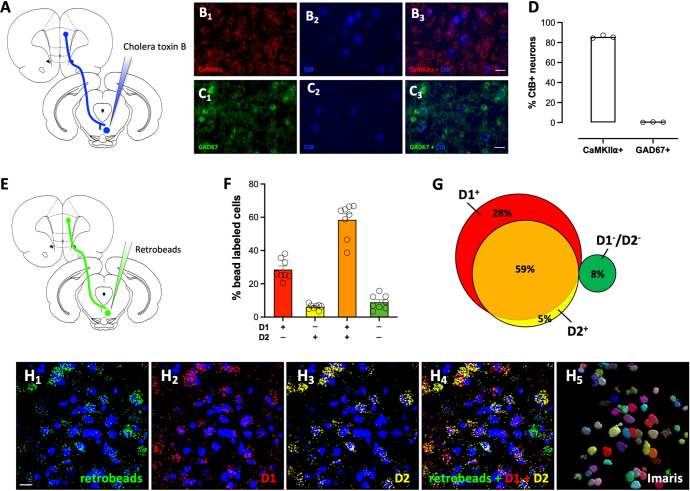
Fig. 2. RMTg-projecting dmPFC neurons express glutamatergic markers and are positive for D1 and D2 receptor mRNA.
A Rats were injected with the retrograde tracer, cholera toxin B (CtB), into the RMTg and slices prepared for dual immunofluorescence. Representative dmPFC images co-labeled for the B1–3 glutamatergic marker CaMKIIα (red) and CtB (blue) and the C1–3 GABAergic marker GAD67 (green) and CtB (blue). Scale bar = 25 μm. D Quantification of co-labeling reveals that RMTg-projecting neurons are CaMKIIα+. E For in-situ hybridization, fluorescent retrobeads were injected into the RMTg and slices processed using RNAScope. F, G Quantification of D1 and D2 mRNA labeling in retrobead+ cells revealed that most RMTg-projecting dmPFC neurons express transcript for both D1 and D2 receptors. H1–5 Representative dmPFC images co-labeled with retrobeads (green), D1 mRNA (red), and D2 mRNA (yellow), as well as an image of Imaris rendered 3D soma, which was used for defining neuronal labeling of the bead and mRNA transcripts. Scale bar = 20 μm.

