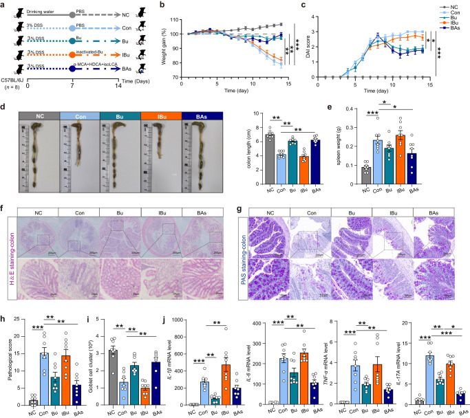
Fig. 5. The mixed bile acids attenuated symptoms in the DSS-induced colitis mouse model.
a Schematic of the experimental design. Mice (female, n = 8 per group) were given 3.0% DSS for 14 d and treated with PBS, Bu, inactivated Bu, or mixed bile acids from d 7 to d 14. b Changes in body weights during the experiments. c DAI scores during the experiments. d A representative image of colon tissue from each group was provided, and the colon length was recorded (n = 8). e The spleen weight of each group (n = 8). f Representative microscopic image of H&E staining of mouse colon tissue (Scale bars = 200 μm) and corresponding local high magnification images (Scale bars = 20 μm). g Representative microscopic image of PAS staining of mouse colon tissue (Scale bars = 200 μm) and corresponding local high magnification images (Scale bars = 100 μm). Histological scores (h) and goblet cell count (i) of the DSS-induced colitis were evaluated (n = 8). j qPCR analysis showing the mRNA expression of IL-1β, IL-6, TNF-α, and IL17A in colon tissues (n = 8). Data were analyzed using one-way ANOVA with Tukey’s test and expressed as the means ± SEM. *p < 0.05, **p < 0.01, ***p < 0.001.

