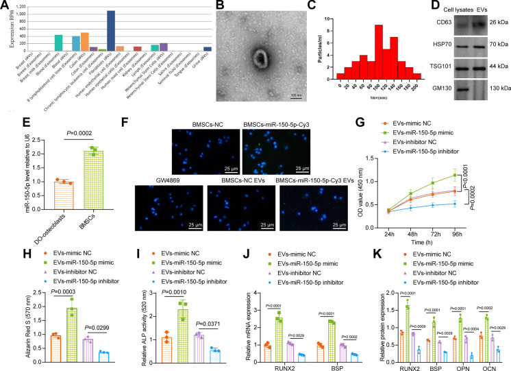
Fig. 2.
BMSC-EV-miR-150-5p stimulates the proliferation and maturation of osteoblasts. A Expression of miR-150-5p in MSC-EVs predicted by the EVmiRNA website (http://bioinfo.life.hust.edu.cn/EVmiRNA#!/). B Morphological characterization of the isolated EVs observed using a TEM. C The size distribution of the isolated EVs analyzed by nanoparticle tracking analysis. D Immunoblotting of EV surface makers CD63, HSP70, and TSG101 along with negative EV marker GM130 in the isolated EVs. E miR-150-5p expression in osteoblasts isolated from DO rats and BMSCs determined by RT-qPCR. F Cy3-labeled green fluorescence signal observed under a fluorescence microscope. G Osteoblast viability of osteoblasts co-cultured with EV-miR-150-5p mimic or EV-miR-150-5p inhibitor measured by CCK-8 assay. H Alizarin red S staining of mineralized nodules in osteoblasts co-cultured with EV-miR-150-5p mimic or EV-miR-150-5p inhibitor. I ALP staining of ALP activity in osteoblasts co-cultured with EV-miR-150-5p mimic or EV-miR-150-5p inhibitor. J mRNA expression of RUNX2 and BSP in osteoblasts co-cultured with EV-miR-150-5p mimic or EV-miR-150-5p inhibitor determined by RT-qPCR. K Immunoblotting of RUNX2, BSP, OPN, and OCN proteins in osteoblasts co-cultured with EV-miR-150-5p mimic or EV-miR-150-5p inhibitor. * p < 0.05. The cell experiment was repeated 3 times independently

