
Figure 4. CD169+ macrophage infection supports systemic dissemination.
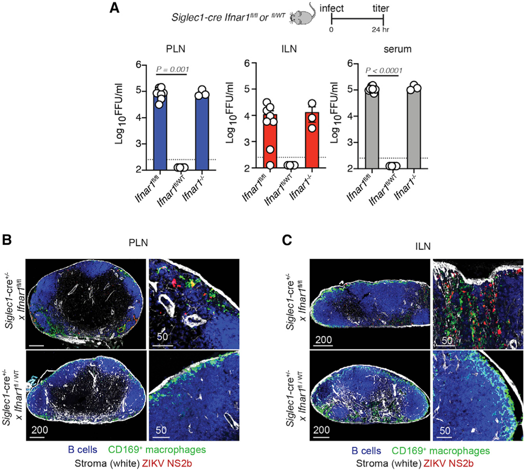
(A) Viral titers (FFUs per milliliter) in the PLN (left, blue bars), ILN (center, red bars), and serum (right, gray bars) harvested 24 h p.i. from Siglec1-cre Ifnarfl/fl (homozygous Ifnar1 knockout in CD169+ cells [cKO]) mice, Siglec1-cre Ifnarfl/WT (heterozygous knockout in CD169+ cells), and Ifnar1−/− mice with 104 FFUs of ZIKV. The experiment was repeated 3 times with 3–4 mice per group. Results shown are pooled from two independent experiments. Dots represent individual mice (either pooled LNs or separate serum) and the average of technical replicates. Dashed line, LOD for the assay. Values below the LOD are reported as half the LOD (125 FFU/mL).
(B) Confocal images of frozen PLN sections harvested 24 h p.i. from Siglec1-cre Ifnarfl/fl (homozygous cKO) mice (top panels) and Siglec1-cre Ifnarfl/WT (heterozygous cKO mice, bottom panels) stained for B cells (using the B220 Ab, blue), CD169+ macrophages (green), stromal cells (using ERTR7, white), and ZIKV NS2b protein (red). The right panels show higher-magnification images. Scale bars are in micrometers.
(C) As in (B) but showing the ILN instead of PLN.