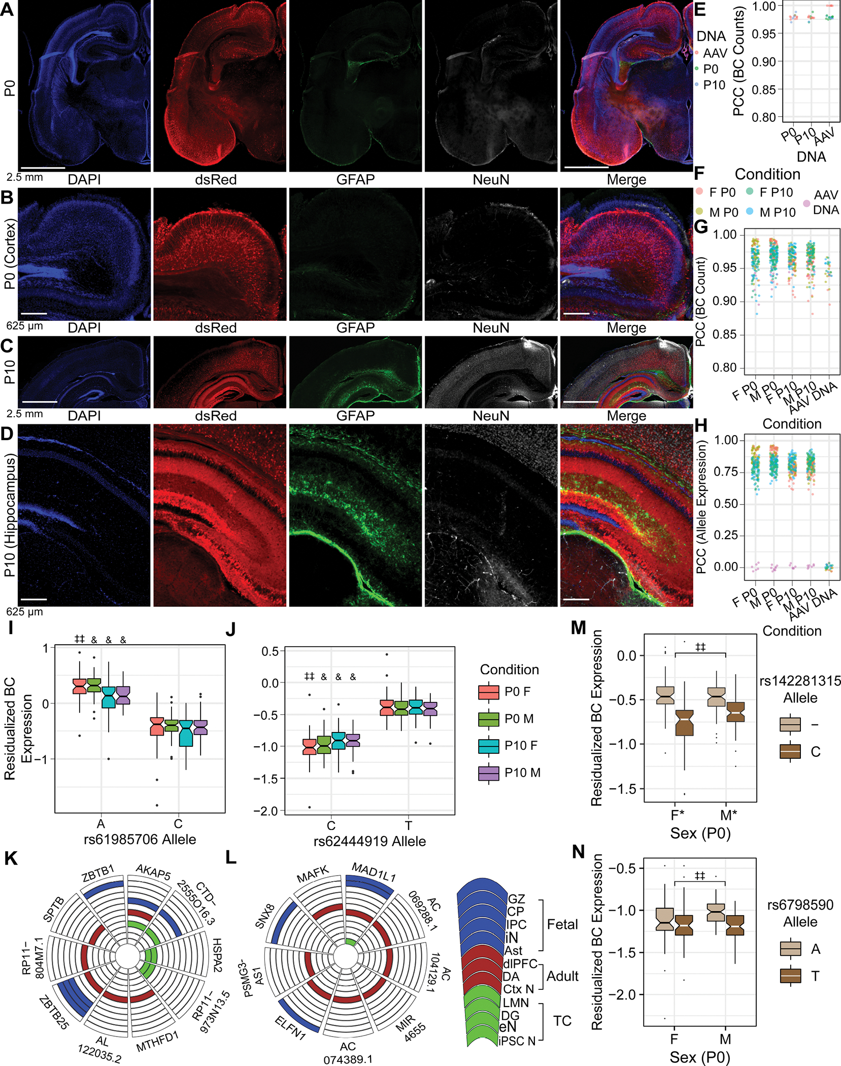
Figure 4. Validating the in utero MPRA delivery method, and identification of rSNPs and sex-interacting rSNPs in the developing brain.

(A, B) IF of P0 brain after E15 MPRA-AAV delivery. (C, D) IF of P10 brain after MPRA-AAV delivery. (E) Comparability of barcode counts in recovered brain DNA and original AAV. (F) Color legend for panels G-H. (G) BC count correlation between samples. (H) Sequence expression correlation between samples. (I, J) rSNPs (I) rs61985706 and (J) rs62444919 showed effects consistent across sexes and ages. (K, L) Putative target genes of the respective rSNPs from Hi-C in human fetal, adult, and cultured neural tissues. (M) Example P0 SxG SNP with comparatively small sex difference in allele effect size. (N) Example P0 SxG SNP with magnitude of sex difference in allelic effect comparable to smaller (female) allelic effect itself. GZ: germinal zone; CP: cortical plate (86); IPC: intermediate progenitor cell; iN: inhibitory neuron (19); Ast: astrocyte (61); dlPFC: dorsolateral prefrontal cortex; DA: dopamine neurons of substantia nigra and ventral tegmental area (65); Ctx N: cortical neuron (60); LMN: lower motor neuron; eN: excitatory neuron (61); iPSC N: iPSC-derived neuron (62). *: pemp-derived FDR < 0.25; **: <0.2; ‡ < 0.15; ‡‡ < 0.1; & < 0.05.
