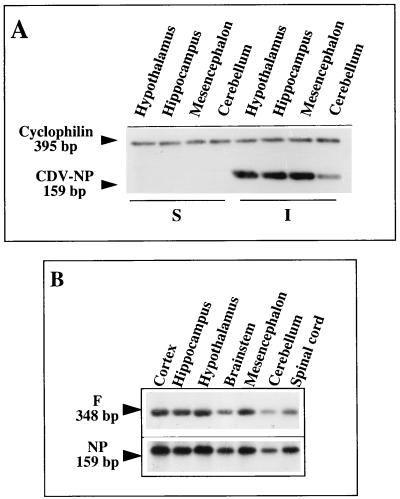
FIG. 3.
Analysis of CDV transcription in the main viral targets in the brain. RT-PCR coamplification of the NP gene and the housekeeping cyclophilin gene (A) and NP and F gene transcripts (B) in various brain structures of infected and noninfected (sham-inoculated) Swiss mice during the acute stage of infection (14 days p.i.) are shown. Visualization of NP (159-bp) and cyclophilin (395-bp) gene amplicons by ethidium bromide staining (A) shows that the same amount of amplified DNA was loaded in each lane and strongly suggests active viral replication in various brain structures, e.g., the hypothalamus. Southern blots (B), using internal probes specific for each viral amplicon (NP and F gene transcripts), allow semiquantification of the PCR products (image analysis, counting excised amplicons in counts per minute) (Tables 2 and 3). The autoradiographs were exposed for 10 min (NP gene amplicon) or 2 h (F gene amplicon), indicating the presence of different amounts of these two viral mRNAs, as expected from their positions on the viral genome. The expected sizes were 159 and 348 bp for the NP and F genes, respectively (Table 1).

