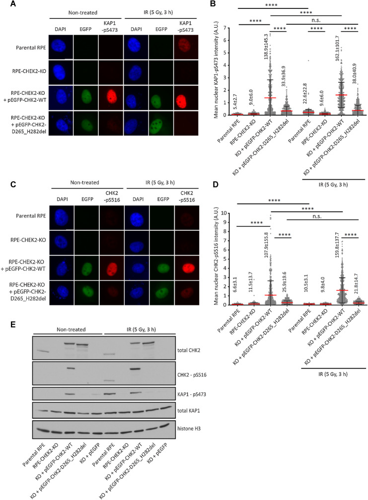
Figure 2.
Validation of KAP1-pS473 and CHK2-pS516 antibodies. A, Parental RPE, RPE1–CHEK2-KO cells or RPE1–CHEK2-KO cells transfected with the wild-type or mutant pEGFP–CHEK2 were left untreated or were exposed to ionizing radiation (5 Gy, 3 hours). After fixation, cells were probed with KAP1-pS473 antibody. Representative images are shown. B, Quantification of A. The mean nuclear intensity of the KAP1-pS473 signal is plotted. Each dot represents one cell; more than 300 cells were analyzed. Red line, error bars and numbers indicate mean ± SDs. Statistical significance was evaluated by the Mann–Whitney test (****, P < 0.0001). A representative experiment is shown from two independent replicates. C, Cells were grown and treated as in A and were probed with CHK2-pS516 antibody. Representative images are shown. D, Quantification of C. The mean nuclear intensity of the CHK2-pS516 signal is plotted. Each dot represents one cell; more than 300 cells were analyzed. Red line, error bars and numbers indicate mean ± SDs. Statistical significance was evaluated by the Mann–Whitney test (****, P < 0.0001). A representative experiment is shown from two independent replicates. E, Cells were grown and treated as in A. Whole-cell lysates were analyzed by immunoblotting with indicated antibodies.

