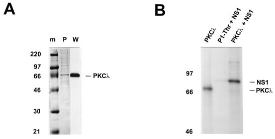
FIG. 2.
Analysis of purified recombinant PKCλ. PKCλ cDNA derived from A9 cells was cloned into pTMHis1, recombined into vaccinia virus, and expressed by coinfection with vTF7-3 in HeLa-S3 cells. The His6-tagged PKCλ was purified from whole-cell extracts on Ni2+-NTA agarose columns. (A) Purified recombinant PKCλ was analyzed by SDS-PAGE (10% polyacrylamide) and detected by Coomassie blue staining (P) or Western blotting with anti-PKCλ (W). Molecular weight markers (m) are indicated on the left, and the apparent migration of PKCλ (70 kDa) is shown on the right. (B) The phosphorylation activity of purified recombinant PKCλ was determined by in vitro kinase assays with [γ-32P]ATP in the absence (PKCλ) or in the presence (PKCλ + NS1) of dephosphorylated NS1. The products were analyzed directly by SDS-PAGE (7% polyacrylamide). P1-Thr, the “kinase-free” P1 fraction used for replication assays, served as a negative control for NS1O phosphorylation (P1-Thr + NS1).

