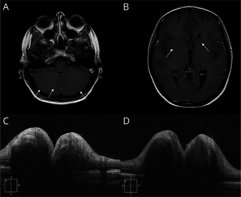
Figure 2. MRI and OCT Features in Patient 2.

Postcontrast axial T1 MRI shows a very subtle radial enhancement and a diffuse venular congestion, both supratentorial and infratentorial, indicated by the white arrows (panels A, B).

Postcontrast axial T1 MRI shows a very subtle radial enhancement and a diffuse venular congestion, both supratentorial and infratentorial, indicated by the white arrows (panels A, B).