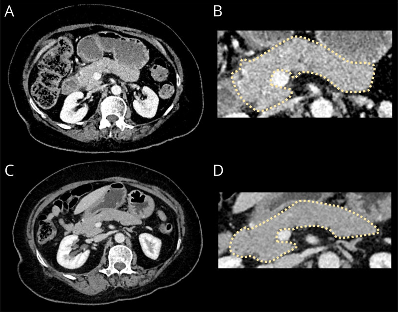
Figure 2. CT of the Chest, Abdomen, and Pelvis Imaging.

Axial contrast-enhanced CT portovenous phase imaging of the abdomen at presentation (A) with a magnified panel of the pancreas (B) demonstrates minimal peripancreatic fat stranding and diffuse enlargement of the pancreas with loss of the definition of the pancreatic clefts causing a “sausage-shape” morphology, typical of IgG4-related disease.6 Repeat CT (C) with a magnified panel (D) 5 months later demonstrates resolution of the fat stranding and reduced pancreatic swelling.
