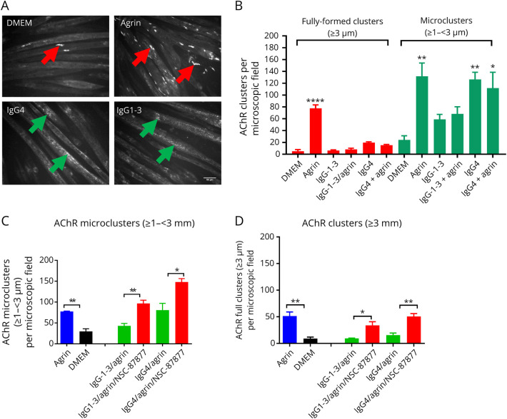
Figure 3. AChR Microclusters Are Reduced in IgG1-3–Treated Myotubes.
Myotubes were exposed to agrin, MuSK-IgG4, and IgG1-3 for 16 hours, and AChR clusters stained with Alexa Flour 594 α-bungarotoxin. Clusters were counted with ImageJ software using different thresholds for cluster length (≥1 – <3 µm for microclusters; ≥3 µm for fully formed clusters). (A) Representative images of AChR clusters. A few fully formed clusters form spontaneously on the myotube surface in DMEM or IgG4-Abs but, as expected, are increased substantially after agrin exposure (red arrows). By contrast, only AChR microclusters (green arrows) were present in myotubes treated with IgG1-3 or IgG4 MuSK-Abs. In this case, fully formed clusters were rarely detectable. (B) However, the results of 3 experiments showed that the AChR microclusters were reduced by approximately 50% in the presence of IgG1-3 compared with either agrin alone or IgG4-treated cells. Scale bar represents 50 µm. One-Way ANOVA with multiple comparisons against DMEM. (C) SHP2 inhibition by NSC-87877 increased the number of AChR microclusters to levels comparable or greater than agrin alone and (D) also the numbers of fully formed clusters. Two-sided t tests for comparisons at each IgG concentration. One-Way ANOVA with multiple comparisons against DMEM. Mean + SEMs are shown for all experiments.

