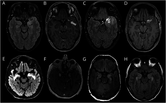
Figure 5. AE Mimic Examples (Brain MRI).
(A–D) Glioblastoma multiforme (GBM): 47-year-old female patient with subacute working memory deficits and new-onset focal seizures. Left temporal hyperintensities on T2/FLAIR images (A) with subtle left temporal leptomeningeal enhancement (B). Brain MRI after 6 months showed progression of T2/FLAIR hyperintense left temporal lesion (C) and enhancement (D). (E–F) CNS Whipple disease: 69-year-old male patient with rapidly progressive dementia and diarrhea. Bilateral mesiotemporal hyperintensities on T2/FLAIR images (E) and parenchymal enhancement in corresponding regions. (This patient was also published elsewhere by Kloek et al.48). (G) Neurofibromatosis type 1 (NF-1): 52-year-old male patient with a chronic course focal epilepsy and cognitive decline. Bilateral mesiotemporal T2/FLAIR hyperintensities, showing no progression for approximately 10 years, regarded as CNS lesion due to NF-1.49 (H) 3,4-Methyl enedioxy methamphetamine (MDMA) intoxication: 27-year-old male patient with acute amnestic syndrome. No seizures were observed, and hippocampal damage was probably causes by direct MDMA-neurotoxicity, as described earlier.50

