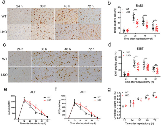
Figure 1.

Loss of MSL1 impairs liver regeneration after partial hepatectomy (PH). a) BrdU staining of liver tissues from WT and LKO mice at 24–72 h after PH. Scale bar, 50 µm. b) BrdU‐positive cell count at 24–72 h after PH, n = 4–5 mice per group, 5 fields (215 × 325 µm2) quantified/animal. c) Ki67 staining of liver tissues from WT and LKO mice at 24–72 h after PH. Scale bar, 50 µm. d) Ki67‐positive cell count at 24–72 h after PH, n = 4–5 mice per group, 5 fields (215 × 325 µm2) quantified/animal. e,f) Serum alanine aminotransferase (ALT) (e) and aspartate aminotransferase (AST) (f) levels in WT and LKO mice at 24–72 h after PH (n = 4 mice per group). g) Liver‐to‐body weight ratio of WT and LKO mice after PH (n = 4–7 mice per group). The data were expressed as means ± SD. Significant difference was presented at the level of * p < 0.05, ** p < 0.01, and *** p < 0.001 by two‐tailed Student's t‐test.
