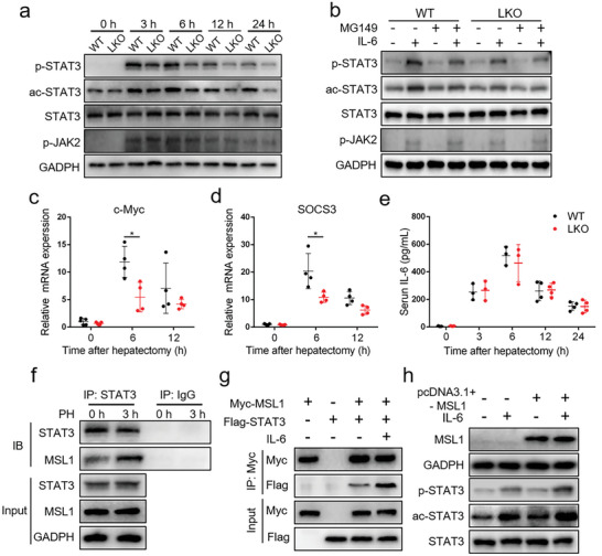
Figure 2.

MSL1 enhances STAT3 transcriptional activity by interacting with STAT3 during liver regeneration. a) Immunoblot analysis of liver tissue lysates from WT and LKO mice at 24–72 h after partial hepatectomy (PH) using the indicated antibodies. b) Immunoblot analysis of primary mouse hepatocytes from WT and LKO mice treated with 50 × 10−6 m MG149 for 24 h with or without 30‐min 10 ng mL−1 IL‐6 treatment using the indicated antibodies. c,d) qRT‐PCR analysis of c‐Myc (c) and SOCS3 (d) mRNA expression in the regenerating liver (n = 4–5 mice per group). e) ELISA analysis of serum IL‐6 level in WT and LKO mice at 24–72 h after PH (n = 3–4 mice per group). f) Coimmunoprecipitation analysis of MSL1 and STAT3 in liver tissues from WT mice. g) HEK293T cells were transfected with Myc‐MSL1‐encoding plasmids and with or without Flag‐STAT3 plasmids, followed by culturing for 24 h without or with 10 ng mL−1 IL‐6 for 30 min. Coimmunoprecipitation analysis was performed. h) Hep1‐6 cells were transfected with plasmids for MSL1 or control vector for 24 h and treated without or with 10 ng mL−1 IL‐6 for 30 min. Immunoblot analysis using the indicated antibodies. The data were expressed as means ± SD. Significant difference was presented at the level of *p < 0.05 by two‐tailed Student's t‐test.
