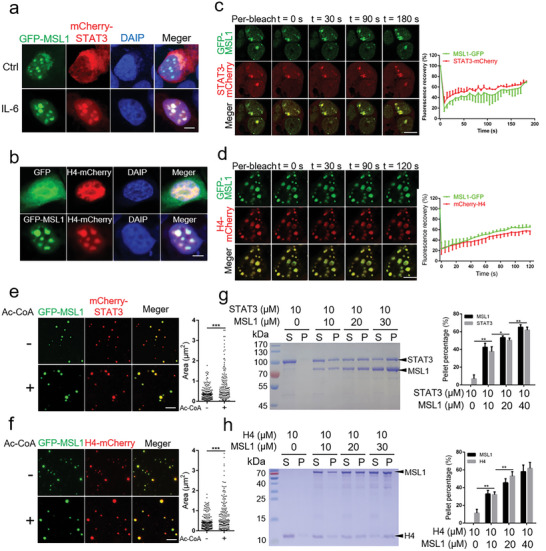
Figure 5.

MSL1 compartmentalizes STAT3 or H4 in phase‐separation condensates to promote the acetylation of STAT3 or H4. a) Confocal images of condensates in fixed HEK293T cells transfected with GFP‐MSL1‐ and mCherry‐STAT3‐encoding plasmids for 24 h without or with 10 ng mL−1 IL‐6 for 30 min. Scale bar, 5 µm. b) Confocal images of phase condensates in fixed HEK293T cells transfected with H4‐mCherry‐encoding plasmid with GFP‐ or GFP‐MSL1‐encoding plasmid transfection. Scale bar, 5 µm. c,d) Fluorescence recovery after photo bleach (FRAP) assays and quantification of condensates in live HEK293T cells transfected with GFP‐MSL1 and mCherry‐STAT3 (c) or H4‐mCherry (d). Data are representative of three independent FRAP events. Scale bar, 5 µm. e,f) Confocal images of GFP‐MSL1 with mCherry‐STAT3 (e) or H4‐mCherry (f) in vitro with or without acetyl‐coenzyme A (Ac‐CoA) (500 × 10−6 m). n = 3 fields (60 × 60 µm2). Scale bar, 5 µm. g,h) SDS–PAGE assay of MSL1 and STAT3 condensates (g) or MSL1 and H4 condensates (h) recovered from the supernatant (S) and pellet (P). Proteins were added at the indicated concentrations. Quantification results of protein fraction recovered from pellets in the sedimentation assays are presented in the right panel. The sedimentation experiments were conducted in triplicates. The data were expressed as means ± SD. Significant difference was presented at the level of * p < 0.05, ** p < 0.01, and *** p < 0.001 by two‐tailed Student's t‐test.
