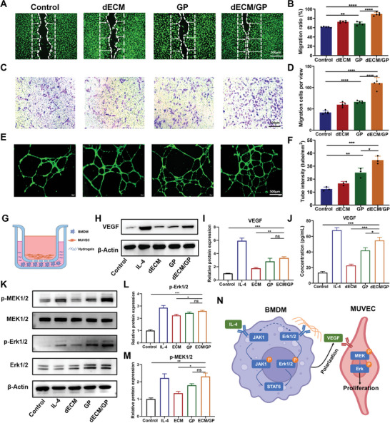
Figure 4.

dECM/GP promoted migration, tube formation and proliferation of endothelial cells. A,B) Migration of HUVECs cocultured with medium or dECM, GP or dECM/GP hydrogels (n = 5). C,D) Representative images and quantification of transwell assay for HUVECs at 24 h (n = 5). E,F) Representative images and quantification of tube formation of HUVECs (n = 3). G) A crosstalk system was established by seeding BMDMs and hydrogels on the bottom and MUVECs on the inserted chamber. H) Western blot analysis of VEGF expression by treated BMDMs. I) Quantitative expression of VEGF (n = 3). J) ELISA of VEGF from supernate of BMDMs’ culture medium (n = 3). K) Western blot of p‐MEK1/2 and p‐Erk1/2. L,M) Quantitative expression of p‐MEK1/2 and p‐Erk1/2 (n = 3). N) Schematic diagram of dECM/GP for proliferation of endothelial cells.
