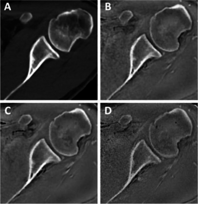
Fig. 6.
Four different axial images of the shoulder demonstrate excellent imaging of the bony structures: A CT; B ZTE MRI 1.0 mm; C ZTE MRI .8 mm; D ZTE MRI .7 mm (from de Mello RAF, Ma YJ, Ashir A, Jerban S, Hoenecke H, Carl M, et al. Three-Dimensional Zero Echo Time Magnetic Resonance Imaging Versus 3-Dimensional Computed Tomography for Glenoid Bone Assessment. Arthroscopy. 2020;36(9):2391–400. Printed with permission)

