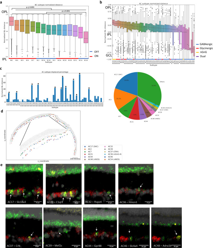
Fig. 4. Laminar organization of neuronal subtypes in the retina.
a Boxplot of bipolar subtype position in the normalized INL length. BC subtypes showed overlapping, yet distinct positioning patterns (ON types in red, and OFF types in blue). RBCs were positioned most apically compared with other subtypes (p-value < 0.001, pairwise t-test, n = 10), whereas BC1B showed significant basal positioning against other subtypes (p-value < 0.001). OFF and ON BC subtypes are marked with blue and red bars, respectively. b Boxplot of amacrine subtype position in the normalized INL length. Most AC subtypes showed distinct positioning within the bottom half of the INL (GABAergic in blue, glycinergic in red, nGnG in yellow, and dual in purple). Displaced AC subtypes showed an increased distribution of cells in the GCL. Glycinergic subtypes showed general apical positioning, whereas GABAergic subtypes showed more basal positioning. 3 nGnG subtypes were the most apically positioned subtypes. GABAergic, glycinergic, nGnG, and dual subtypes are marked with blue, red, yellow, and purple bars, respectively. In the box plots, the bounds of the boxes represent 25 to 75% percentiles with the center lines showing the median. The whiskers extend 1.5 times beyond inter-quartile ranges. Individual points determined to be outliers are visualized outside of the whiskers. c Bar plot of displacement proportion in each amacrine subtype and a pie chart of displaced subtype composition in the GCL. Twelve AC subtypes showed significant displacement using a permutation test by shifting the subtype label for 100 times (p-value < 0.05, n = 59 sections across 10 independent experiments) and made up about 70% of all ACs in the GCL. d Tissue plot of displaced amacrine subtypes. Displaced AC subtypes showed distribution across both INL and GCL. e In-situ hybridization images of displaced AC subtype markers in the ganglion cell layer (n = 2). Specific markers against nine displaced AC subtypes were profiled in conjunction with pan-AC marker Slc32a1 and pan-RGC marker Slc17a6. Specific subtype markers in yellow showed co-localization with the pan-AC marker in green, but not with the pan-RGC marker in red.

