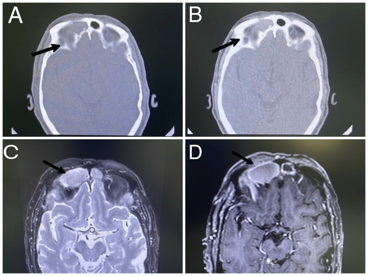
Figure 3. Case 1. Right frontal mucocele in male patient presenting with right eye ptosis, diplopia, and headache. (A) Axial CT scan bone window at level of the frontal sinus, showing expansile mass in right frontal sinus with bone thinning in both anterior and posterior table; (B) Soft tissue window; (C) T1 weighted MRI with gadolinium axial cut showing low signal with peripheral enhancement intensity; (D) T2 FLAIR MRI indicating high signal intensity lesion.
FLAIR: fluid-attenuated inversion recovery

