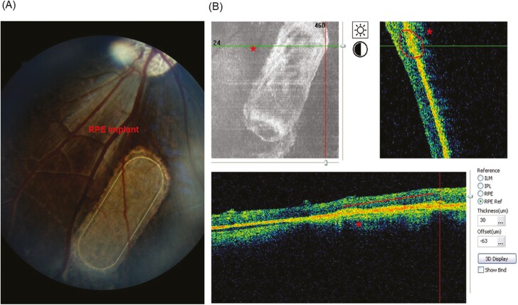
Figure 3.
Non-invasive postoperative examinations of RPE implant by OCT (optical coherent tomography) and fundus photography (unpublished data from our laboratory). (A) Oval nanofibrous implant seeded with cultured porcine primary RPE cells (red arrows) and transplanted into subretinal space of minipigs eye was examined by fundus camera at 8 weeks after implantation and (B) by OCT at 4 weeks after implantation. ‘*’, Sub-retinally transplanted RPE implant in the minipig eyes.

