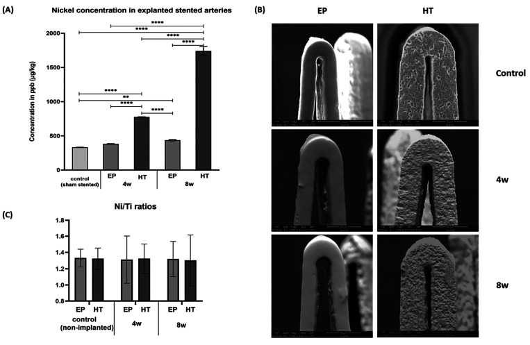
Figure 2.
(A) Nickel levels in explanted sham and stented arteries at 4 and 8 weeks postimplantation. Due to the low concentrations detected, the tissue-digested solution from all samples was collected and pooled (n = 3 per stent type and time point) for more accurate estimates. (B) Representative SEM images of control vs tested (implanted) stents. (C) Ni/Ti ratios of each stent type before and post the 4- and 8-week implantation time. (** indicates p < 0.01 and *** p < 0.001)

