Abstract
Asymptomatic chronic retinal vein occlusion that occurs in chronic simple glaucoma is described. The condition is characterized by marked elevation of retinal vein pressure with collateral vessels and vein loops at the optic disc in cases of central vein occlusion, or retinal veno-venous anastomoses along a horizontal line temporal and nasal to the disc in hemisphere vein occlusion. No patient had visible arterial changes, capillary closure, fluorescein leakage, or haemorrhages. The vein occlusion was not limited to "end stage" glaucoma. The role of increased intraocular pressure and glaucomatous enlargement of the optic cup with retinal vein distortion in the pathogenesis of the condition was stressed. Follow-up of these patients revealed persistence of the retinal vein occlusion shown by elevated retinal vein pressures. This would reduce effective perfusion of the inner retina and optic disc and may affect the long-term visual prognosis.
Full text
PDF
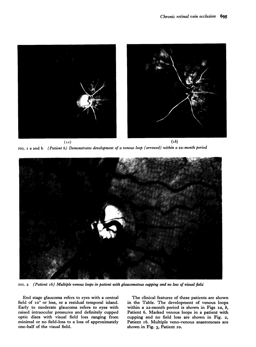
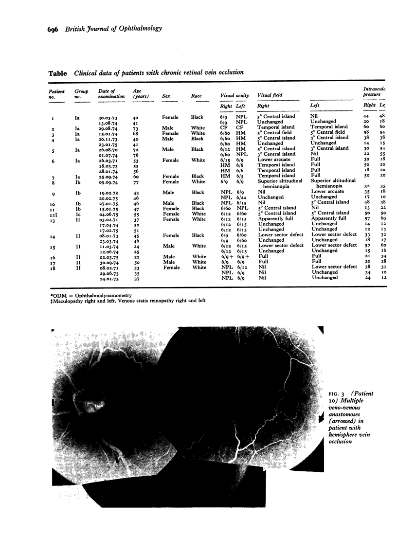
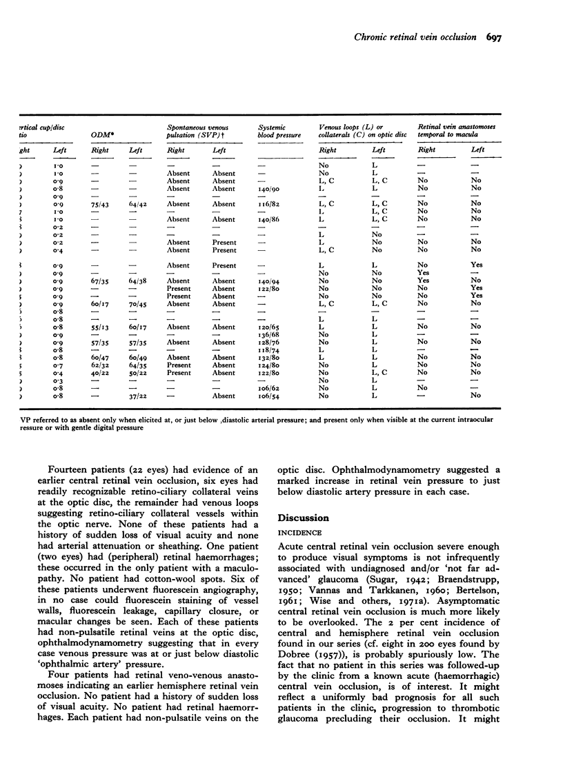
Images in this article
Selected References
These references are in PubMed. This may not be the complete list of references from this article.
- DOBREE J. H. Venous obstruction and neovascularization at the disc in chronic glaucoma. Trans Opthal Soc U K. 1957;77:229–238. [PubMed] [Google Scholar]
- Hayreh S. S. Occlusion of the central retinal vessels. Br J Ophthalmol. 1965 Dec;49(12):626–645. doi: 10.1136/bjo.49.12.626. [DOI] [PMC free article] [PubMed] [Google Scholar]
- VANNAS S., TARKKANEN A. Retinal vein occlusion and glaucoma. Tonographic study of the incidence of glaucoma and of its prognostic significance. Br J Ophthalmol. 1960 Oct;44:583–589. doi: 10.1136/bjo.44.10.583. [DOI] [PMC free article] [PubMed] [Google Scholar]


