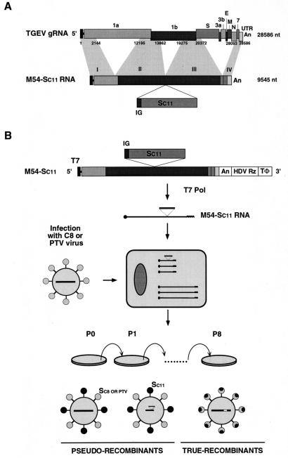
FIG. 6.
Diagram of the pseudorecombinant and true recombinant virus isolation protocol. (A) Structure of the M54 minigenome indicating the sequence position where the S gene from the enteric C11 isolate was cloned. Letters and numbers above the top bar indicate the TGEV ORFs. Numbers below this bar indicate the nucleotide sequences of the helper virus incorporated into the M54 minigenome. Numbers above the second bar indicate the four sequence domains that were linked during the generation of the minigenome. Numbers to the right of the bars indicate sizes of the genomes in nucleotides. gRNA, genomic RNA. IG, intergenic sequences preceding the S gene of the C11 isolate, which is identical to that of the PUR46-MAD strain of TGEV. An, poly(A). (B) Generation of recombinants. The S gene from an enteric TGEV (isolate C11) was cloned into minigenome M54, generating the minigenome M54-Sc11 with the structure diagrammatically shown in panel A. This minigenome has been cloned after the T7 promoter (T7) (black box) and preceding the hepatitis delta virus ribozyme (HDV Rz) sequences and the T7 terminator sequences (TΦ). ST cells were infected with C8 or PTV viruses. At 4 to 6 h p.i., cells were electroporated with in vitro-transcribed RNA and the supernatants from these cultures were passaged by using ST cells. The potential pseudorecombinants with the S protein from the respiratory helper viruses (light circles) or from the enteric C11 isolate (dark circles) containing either the genome of the helper virus (large bar) or the minigenome with the S gene of the C11 isolate are diagrammatically represented (bottom left). True recombinants with a chimeric S protein generated by two crossovers between the S gene from the helper virus and the S gene from isolate C11 are also indicated (bottom right).

