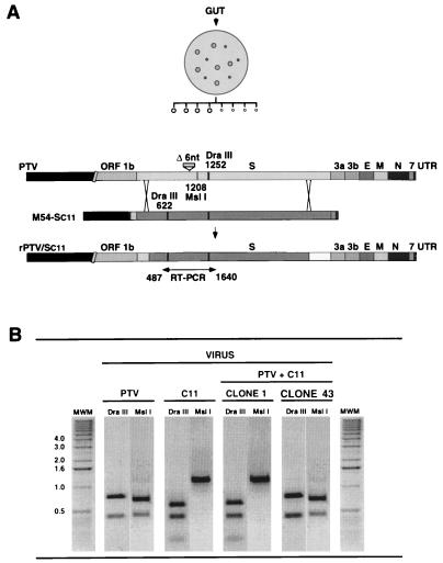
FIG. 8.
Restriction endonuclease analysis of the potential recombinants. (A) A diagram of the procedure followed in the selection of true recombinant and pseudorecombinant viruses growing in the enteric tract is shown. Gut tissue (jejunum), collected from a single pig at 2 d p.i., was homogenized, and lysis plaques were isolated on ST cells. Plaques of two sizes (3- and 1-mm diameter) were observed and cloned twice. Viruses in these plaques were expected to have either the genome of the helper respiratory virus (PTV) or the genome of a true recombinant virus (rPTV/Sc11) formed by a two-crossover event within the S gene (bottom bar). The positions of restriction endonuclease (DraIII and MslI) sites in cDNA, derived by RT-PCR, between nt 487 and 1640 of the S gene are indicated. (B) Prototype restriction endonuclease patterns of cDNAs derived from S genes of the strains PTV and C11 and two isolates (clones 1 and 43) with a restriction endonuclease pattern identical to that of strain C11 or PTV are shown. MWM, molecular weight markers (1-kb DNA ladder; Gibco). Numbers on the left of the MWM lane indicate the sizes of the markers in kilobases.

