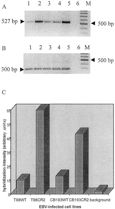
FIG. 2.
Detection of EBV DNA on astrocyte lines 48 h after infection. DNA from each cell line was prepared and used as a template for PCR amplification of a 527-bp fragment of the BamHI W fragment of the EBV genome. (A) PCR amplification products were resolved on a 2% agarose gel and detected by staining with ethidium bromide (inverted picture). A negative control that lacked template DNA (lane 6) and a positive control that used B95-8 DNA as a template (lane 5) were included. Lane 1, T98WT; lane 2, T98CR2; lane 3, CB193WT; lane 4, CB193CR2; M, molecular weight marker (100-bp ladder). (B) The same DNA preparation was amplified for the GAPDH gene by using specific primers. (C) Semiquantification of the EBV DNA PCR signal. After Southern blotting, the membrane was hybridized with a specific 32P-labeled probe. The hybridization signal was analyzed with a phosphorimager and calculated as arbitrary units. The background lane shows the hybridization signal from the negative control. The signal obtained for the positive control was too strong, and the quantification was impossible.

