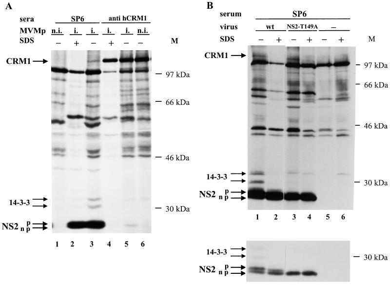
FIG. 5.
Association of CRM1 with the nonphosphorylated forms of NS2 in MVM-infected mouse cell extracts. 35S-labeled extracts were prepared from A9 cells that were mock treated (n.i.) or infected (i.) with either wild-type (wt) MVM (A and B) or mutant (NS2-T149A) MVM (B). Immunoprecipitation reactions were performed with SP6 antiserum (A and B) or anti-hCRM1 antibodies (A) in the presence (+) or absence (−) of SDS and analyzed as described in the legend to Fig. 4. Upper and lower panels in panel B correspond to autoradiograms of the same gel exposed for long and short times, respectively. Arrows to the left point at phosphorylated (p) and nonphosphorylated (np) NS2, 14-3-3, and CRM1 proteins. M, molecular sizes (in kilodaltons) of prestained protein standards.

