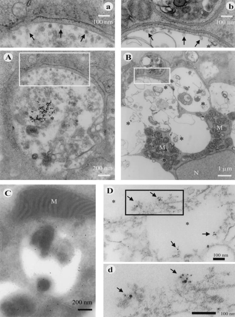
FIG. 3.
Ultrastructure of cytopathic vacuoles in RUB-infected Vero cells. (A) CPVI at 18 h after RUB infection showing endocytosed BSA-gold in the lumen (asterisks) and small spherules at the inner aspect of the vacuolar membrane (enlarged in panel a). (B) General view of a large vacuolar structure at 24 h p.i. containing BSA-gold (asterisks) with numerous mitochondria (M) near the nucleus (N). The enlargement in panel b demonstrates spherules (arrows), BSA-gold (asterisks), and the close proximity of rough ER membranes aligning with the vacuole membrane. (C) Localization of P150 by the cryoimmuno technique to the membrane of a vacuole. Anti-p55 antibody was detected by 10-nm gold–protein A particles. (D) Pre-embedding immunoelectron microscopic image of Triton X-100-treated Vero cells at 48 h p.i. Shown is the intracellular localization of RUB P150 together with metabolically incorporated bromouridine. P150 is labeled with 5-nm gold particles, the bromouridine-RNA is labeled with 10-nm gold particles, and both are visualized best in the enlargement (d). Arrows point to labeled spherule structures. The vacuolar lumen is marked with asterisks, and M stands for mitochondria.

