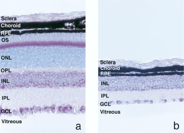
FIG. 1.
Light micrographs of retinas collected from normal (a) and rd (b) mice at 6 weeks of age. Paraffin-embedded sections of eyes were counterstained with toluidine blue. In the rd mouse, the photoreceptor layer that includes the outer segment (OS), the outer nuclear layer (ONL), and the outer plexiform layer (OPL) has degenerated completely. RPE, retinal pigment epithelium; INL, inner nuclear layer; IPL, inner plexiform layer; GCL, ganglion cell layer. Original magnification, ×400.

