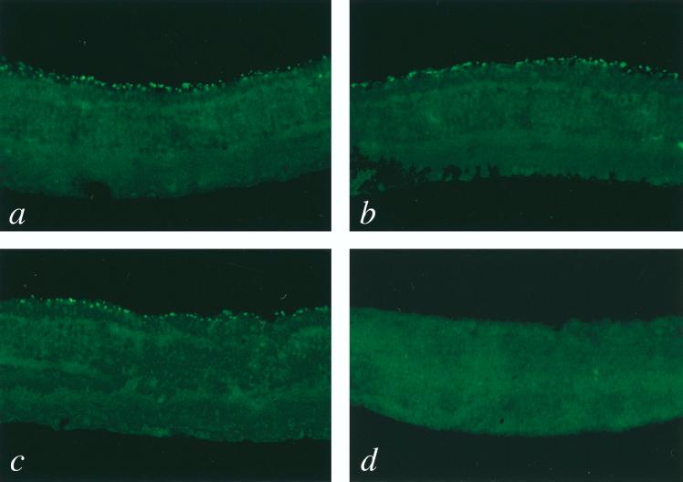
FIG. 3.
Immunohistochemical detection of opsin-positive photoreceptor cells in rd mice following injection of HIV vectors. Mice injected with the Rho-PDEβ vector (a, b, and c) or the Rho-GFP vector (d) were sacrificed at 6 (panels a and d), 12 (panel b), and 24 (panel c) weeks postinjection, and eye sections were prepared as described previously (26). Sections were stained with mouse anti-opsin antibody (kindly provided by C. J. Barnstable) and then with fluorescein isothiocyanate-conjugated donkey anti-mouse immunoglobulin G (Jackson Immunochemicals). Immunofluorescence was detected with a confocal laser scanning microscope (Bio-Rad). Representative confocal microscope images of sections are shown. Opsin-expressing photoreceptor cells (bright green) were seen only in eyes injected with the Rho-PDEβ vector. Original magnification, ×200.

