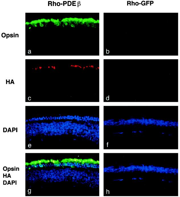
FIG. 4.
Two-color confocal immunofluorescence analysis of rd mouse eyes injected with HIV vectors. Sections of rd mouse eyes obtained 6 weeks after injection of the Rho-PDEβ vector (a, c, e, and g) or the Rho-GFP vector (b, d, f, and h) were stained with mouse anti-opsin antibody and rabbit anti-HA antibody (MBL, Nagoya, Japan). Primary antibodies were detected with fluorescein isothiocyanate-conjugated donkey anti-mouse immunoglobulin G (IgG) (green) (panels a and b) and cyan red-conjugated donkey anti-rabbit IgG (red) (panels c and d) and visualized by confocal laser scanning microscopy. Cell nuclei were counterstained with DAPI (blue) (panels e and f). Double-labeled cells (yellow) demonstrate HA-tagged PDEβ expression in the outer segments of photoreceptor cells (panel g). Original magnification, ×400.

