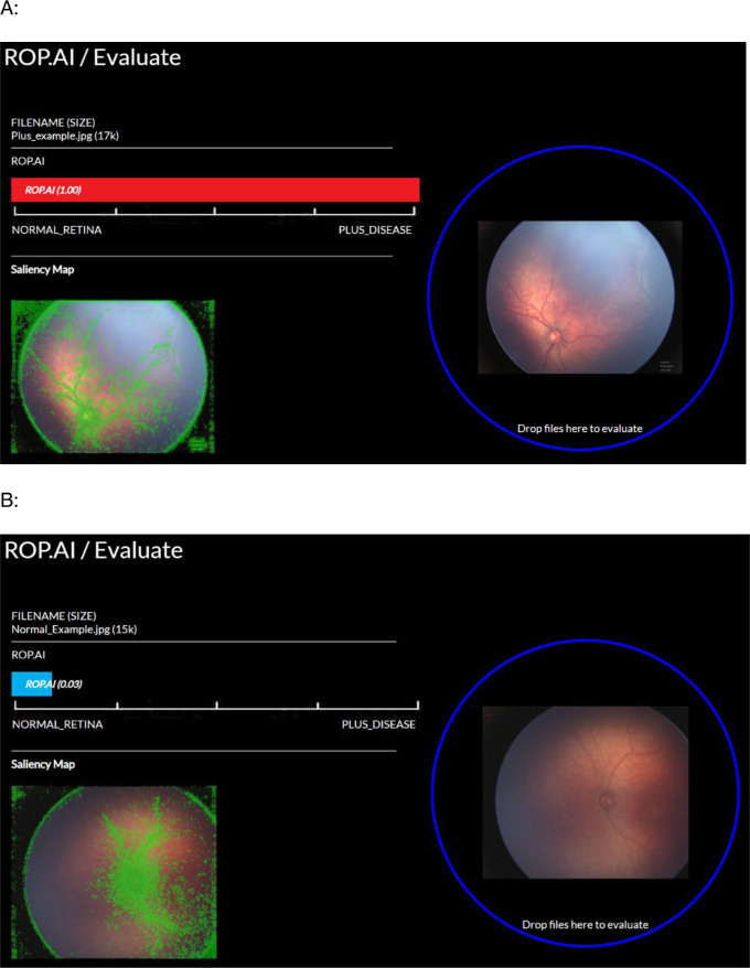
Figure 1.
The ROP.AI platform used to upload retinal images for AI analysis. Corresponding AI evaluation of the retinal image is seen on the bar on the left. An output between 0 and 1 is provided by the AI with a saliency map for each image uploaded seen bottom left. (A) Example analysis of a plus disease image graded by ROP.AI as 1.00 (plus disease). (B) Example analysis of a normal image graded by ROP.AI as 0.03 (normal).

