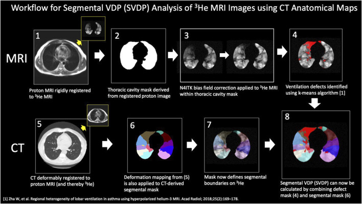
FIGURE 1.
Steps in the analysis workflow to align segmental mask from CT to the 3He MRI ventilation images. The steps are as described in the body of the figure. The segmental VDP (sVDP) was then compared to the corresponding segmental airway fSAD from the parametric response map of the CT scan at the expiratory (FRC) lung volume. Note that the PRM map is inherently registered to CT and so can be readily compared by airway segment with 3He MRI after these analysis steps. Modified from Mummy et al. (2022). Used with permission.

