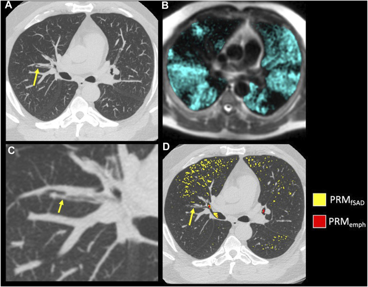
FIGURE 7.
Example of mucus plugging within a moderate asthmatic. (A) A mucus plug (arrow) visualized on inspiratory CT using a MIP of 7 slices. (B) A close-up of the mucus plug (arrow) in (A) (C) HP 3He MRI overlaid on conventional MRI with ventilation defects occurring near and downstream of the mucus plug visualized in (A). (D) PRM maps overlaid on Inspiratory CT in the axial slice of the mucus plug (arrow).

