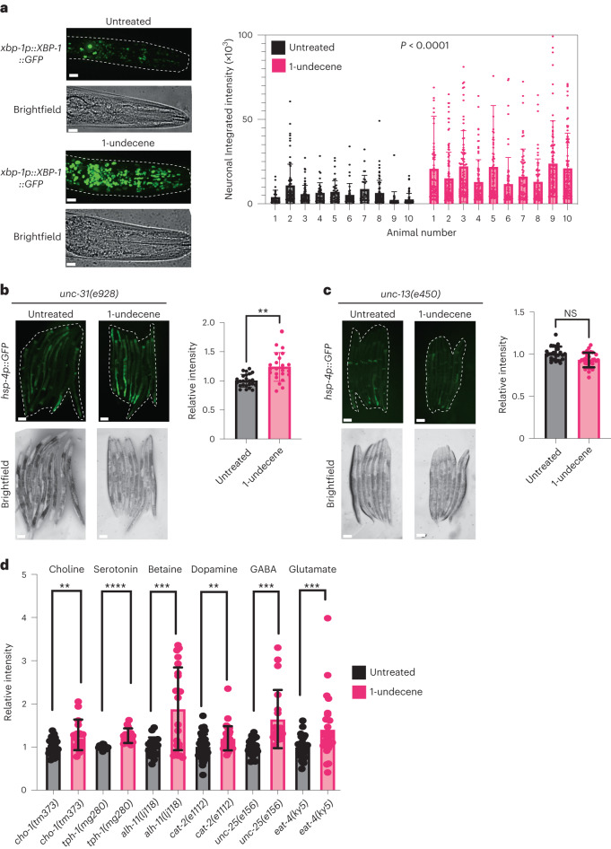
Fig. 2. Neuronal signaling is required for downstream UPRER activation by 1-undecene exposure.
a, Representative image and quantification of GFP+ cells in the head of worms expressing an xbp-1p::xbp-1::GFP transgene with or without exposure to 1-undecene for 8 h. This experiment was repeated three times with 10 worms per group. Scale bars, 10 μm. Graphs show mean ± s.d. Significance was calculated by unpaired Student’s t-test. b,c, Representative fluorescence microscopy images and quantification of hsp-4p::GFP fluorescence in unc-31(e928) (b) and unc-13(e450) (c) with or without exposure to 1-undecene odor for 12 h. These experiments were repeated three times (n = 24 and 23 animals for b and n = 23 and 30 animals for c). Scale bars, 200 μm. Graphs show mean ± s.d. NS, not significant and **P < 0.01 (two-tailed unpaired Student’s t-test) for b and c. d, Quantification of hsp-4p::GFP fluorescence in cho-1(tm373), tph-1(mg280), alh-1(ij118), cat-2(e1112), unc-25(e156) and eat-4(ky5) mutants. Intensity was normalized to untreated animals for each mutant strain. These experiments were repeated three times (n = 20, 16, 12, 15, 18, 22, 44, 39, 21, 20, 36 and 28). Graphs show mean ± s.d. **P < 0.01, ***P < 0.01 and ****P < 0.001 (two-tailed unpaired Student’s t-test comparison between untreated and 1-undecene). Precise P values are provided in Source Data.

