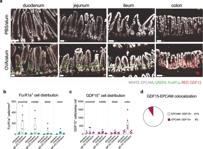
Extended Data Fig. 9. GDF15 originates from colonic epithelial cells.
a, Fcεr1a (green), EPCAM (grey), and GDF15 (red) transcripts across intestinal tissues in OVA/alum BALB/c sensitized and control mice by RNAScope. Scale bars = 50 µm. b-c, Analysis of intestinal distribution of FcεR1 expressing cells (b) and GDF15 expressing cells (c) from control and allergic sensitized WT or IgE KO mice (n = 4 WT control, 5 WT allergic, 4 IgE KO allergic per group). Quantification was performed after RNAscope technique. d, Colocalization analysis of GDF15 expressing colonic cells and cells expressing the epithelial cell marker, EPCAM utilizing sum of all WT 5 allergic mice. Graphs show mean ± s.e.m. *p ≤ 0.05, **p ≤ 0.01, ***p ≤ 0.001. Two-way ANOVA by Genotype/Sensitization Status and Anatomical Region. Representative of two independent experiments.

