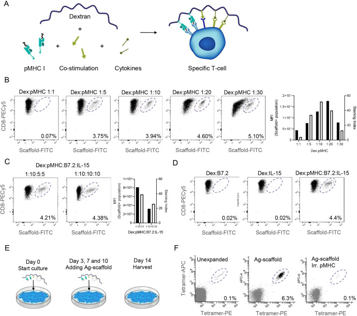
Figure 1.
Ag-scaffolds bind to specific T-cells via attached pMHC I and delivers co-attached co-stimulation and cytokines. (A) Schematic drawing of an Ag-scaffold comprizing a dextran backbone to which MHC class I molecules carrying peptide (pMHC), co-stimulatory molecules and cytokines can be attached. The Ag-scaffold bind to T-cells via pMHC-TCR-interaction and forms an artificial immunological synapse. (B) Binding of FITC-labeled dextran-backbone with varying amounts of pMHCs, to HLA-A0101 CMV pp65 YSE-specific T-cells (~4%). Ratios were compared in median fluorescent intensity (MFI in black) and staining index (SI in white). CD8+ T-cells were labeled with PECy5. (C) T-cell binding of a 1:10 dextran:pMHC in combination with either 5:5 or 10:10 of B7.2:IL-15. (D) Ag-scaffolds comprizing only dextran:B7.2 (1:30) or dextran:IL-15 (1:30) could not direct T-cell binding. (E) Schematic representation of a 2 week Ag-expansion where Ag-scaffold and fresh media is supplemented to the culture on day 0, 3, 7 and 10. (F) Only Ag-scaffolds (dextran:pMHC:B7.2:IL-15) containing HLA-A0101 carrying the Influenza BP VSD-peptide and not an irrelevant pMHC, could expand a population of HLA-A0101 Influenza BP VSD-specific T-cells from PBMCs from a healthy donor. Specific cells were stained with a PE/APC-tetramer. Ag-scaffolds, antigen-presenting scaffold; APC, Allophycocyanin; BP, polymerase basic protein, CMV, cytomegalovirus, dex, dextran; FITC, Fluorescein isothiocyanate; HLA, human leukocyte antigen; IL, interleukin; MHC, Major Histocompatibility Complex; PBMCs, peripheral blood mononuclear cells; PE, R-phycoerythrin; TCR, T-cell receptor; VSD, the first three aminoacids of the peptide.

