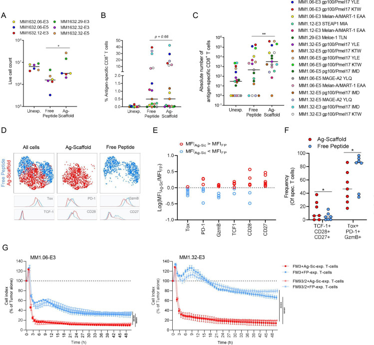
Figure 4.
Ag-scaffolds efficiently expand tumor-specific T-cells and render a multifunctional T-cell product. (A) Number of viable cells at culture initiation (Unexp.) and following 2-week expansion with either dextran:pMHC:IL-2:IL-21 Ag-scaffold or free peptide-based expansion, adding specific peptide, IL-2 and IL-21 directly to the media, targeting melanoma shared antigens in six patients with metastatic melanoma samples (PBMC). (B) Post-expansion frequency and (C) number of tumor-specific T-cells (seven specificities) from 17 expansions from six patients with metastatic melanoma samples, with either dextran:pMHC:IL-2:IL-21 Ag-scaffold or free peptide-based expansion adding specific peptide, IL-2 and IL-21 to the media. (D) UMAP plot of HLA-A0201 gp100/Pmel17 YLE-specific T-cells expanded from patient MM1.06-E5 with either Ag-scaffold or free peptide. Specific T-cells were identified by tetramer-staining and stained with an antibody panel including markers of self-renewing (TCF-1, CD28, CD27) and terminally differentiated/exhausted (Tox, PD-1, GzmB) T-cells. The expression of these markers on Ag-scaffold (red) and free peptide (blue)-expanded T-cells were compared in MFI histograms. (E) Extended phenotypic comparison between Ag-scaffold and free peptide-expanded T-cells (Log(MFIAg-Sc/MFIFP) across two different tumor-specific T-cell populations expanded from four patient samples. (F) Frequency of TCF-1+CD28+CD27+ and PD-1+GzmB+Tox+ T-cells out of antigen-specific T-cells expanded with either Ag-scaffold and free peptide. (G) Survival of A0201-positive FM3 and FM93/2 melanoma tumor cells (Cell Index as a relative measure of impedance presented as % of tumor alone) on co-culture with Ag-scaffold (light/dark red) or free peptide-expanded (light/dark blue) tumor-specific T-cells from MM1.06- and 32-E3 in a 4:1 E:T ratio. Duplicate-means were compared in a paired, non-parametric Wilcoxon test and p<0.05 and p<0.01 are indicated as * and **, respectively. Ag-scaffolds, antigen-presenting scaffold; GzmB, granzyme B; HLA, human leukocyte antigen, IL, interleukin; MFI, mean fluorescence intensity; PBMCs, peripheral blood mononuclear cells; PD-1, Programmed cell death protein 1; pMHC, peptide-MHC; Tox, Thymocyte selection-associated high mobility group box protein; UMAP, uniform manifold approximation and projection.

