Figure 5.
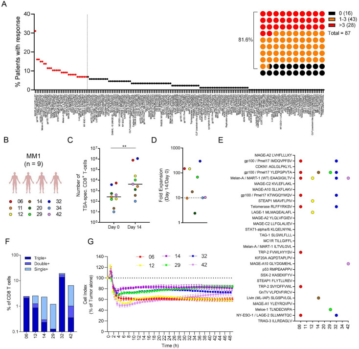
Expansion of tumor-specific T-cells using a multitargeting shared melanoma Ag-scaffold. (A) Percentage of patients showing reactivity towards the library of HLA-A0201-restricted TSAs. Marked in red are the top 30 most commonly recognized shared melanoma antigens among the evaluated patients. To the right, a schematic representation showing that 43/87 (~49%) patients showed reactivity against 1–3 of the selected 30 shared melanoma antigens and 28/87 (~32%) showed reactivity against >3. (B) PBMCs from a cohort of nine patients with metastatic melanoma (MM1) were expanded with the multitargeting shared melanoma Ag-scaffold, comprizing 30 Ag-scaffolds carrying the 30 selected TSAs on HLA-A0201. (C) Number of TSA-specific T-cells pre-expansion (Day 0) and post-expansion (Day 14) were compared in a paired, non-parametric Wilcoxon test and p<0.01 is indicated as **. (D) Fold expansion (FE = Day 14/Day0). (E) Post-expansion T-cell populations among the 30 shared melanoma antigens. (F) Frequency of Triple+, Double+ and Single+for IFN-γ, TNF-α and CD107a+ out of the total CD8+ T-cell population on challenge with cognate-antigen (identified in Figure 6E) in the six patients with successful expansion of melanoma-specific T-cells. (G) Survival of HLA-A0201-positive FM93/2 melanoma tumor cells (Cell Index as a relative measure of impedance presented as % of tumor alone) when co-cultured with Ag-scaffold-expanded T-cells from the six patients with melanoma in a 4:1 E:T ratio. Ag-scaffolds, antigen-presenting scaffold; E:T, effector:target; HLA, human leukocyte antigen; IFN, Interferon; PBMCs, peripheral blood mononuclear cells; TNF, tumor necrosis factor; TSA, tumor shared antigen.

