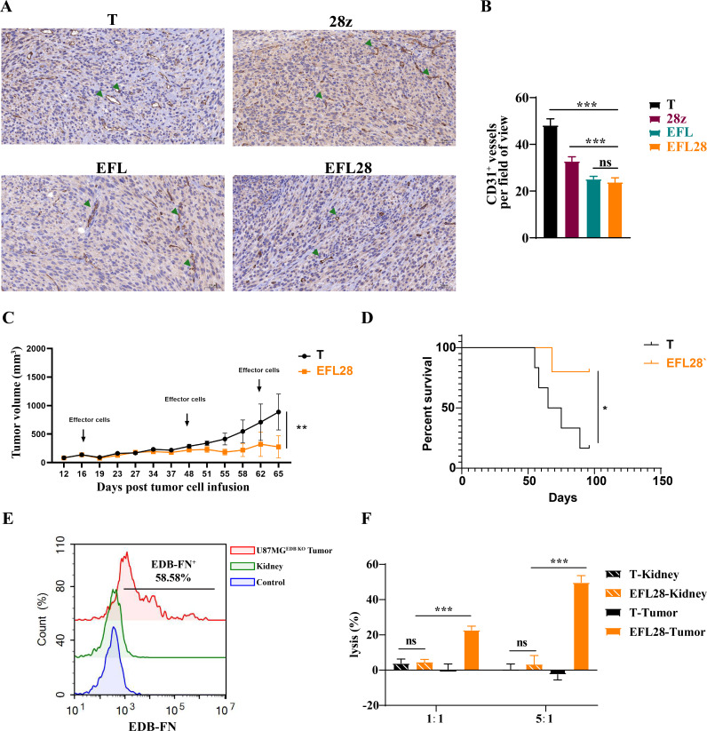
Figure 4.
EFL28 rTCR-CAR T cells reduce tumor blood vessels. (A) 107 CAR T cells or T cells were injected into the tail vein of U87MG tumor-bearing mice, tumor tissues were collected on the 14th day, and tumor-associated vascular endothelial cells were detected by immunohistochemistry using an anti-CD31 antibody. (B) Quantitation of CD31+ vessels. Statistical significance was calculated by one-way analysis of variance (ANOVA) with Bonferroni post hoc test, n=5, ***p<0.001. (C) Tumor growth of different groups. Statistical significance was calculated by two-way ANOVA; mice treated with T cells, n=6; mice treated with EFL28 rTCR-CAR T cells, n=5. **p<0.01 (D) Survival plot using a Kaplan-Meier curve. Statistically significant differences were determined using the log-rank test, *p<0.05. (E) CD31-positive cells were isolated from the kidneys and tumors of U87MGEDB KO tumor-bearing mice. EDB expression in vascular endothelial cells of the kidney or U87MGEDB KO tumors was analyzed by flow cytometry using the L19 antibody. (F) CAR T cells were cytotoxic to EDB-positive cells of tumor origin but not to endothelial cells from the kidney. Statistical significance was calculated by one-way ANOVA with Bonferroni post hoc test, n=3, ***p<0.001. EDB, extra domain B.

