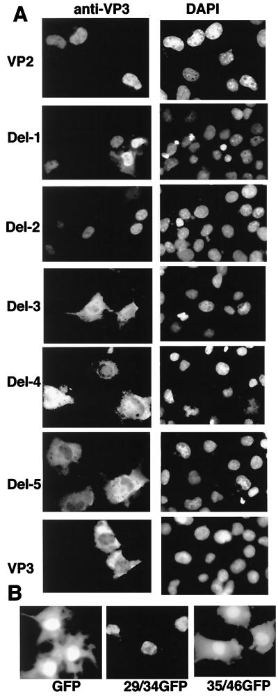
FIG. 2.
NLS activity of the VP2 N-terminal region. (A) Subcellular distribution of the N-terminal deletion mutants of VP2. COS1 cells were transfected with plasmids containing VP2, VP3, and the panel of VP2 deletion mutants. Localization of individual proteins was analyzed by indirect immunofluorescence. Cells were stained with anti-VP3 antibody and subsequently with rhodamine-conjugated rabbit anti-immunoglobulin G. 4′,6-Diamidino-2-phenylindole (DAPI)-stained COS1 cells in the same fields are also shown. (B) NLS activity of the peptide from the VP2 N-terminal region. Fluorescent microscopic images of GFP and GFP-fusion proteins expressed in COS1 cells are shown. 29/34GFP and 35/46GFP indicate the localization of the fusion proteins having the amino acid 29 to 34 and 35 to 46 regions, respectively.

