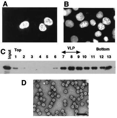
FIG. 3.
VLP formation of NLS-VP3. (A) Subcellular distribution of NLS-VP3. COS1 cells were infected with plasmids containing the NLS-VP3 construct and after 48 h of transfection, the subcellular distribution of NLS-VP3 was analyzed by immunofluorescence staining with anti-VP3 antibody followed by rhodamine-conjugated rabbit anti-immunoglobulin G. (B) DAPI-stained COS1 cells. The image is the same field as shown in panel A. (C) Sucrose density gradient analysis of NLS-VP3. Sf9 cells were infected with a recombinant baculovirus containing NLS-VP3 and the cell extract was subjected to sucrose density gradient to analyze for VLP formation. Each fraction was subjected to SDS-PAGE followed by immunoblotting with anti-VP3 antibody. (D) Electron microscopy. VLPs were purified by CsCl density gradient ultracentrifugation and analyzed by electron microscopy with negative staining. Bar, 100 nm.

