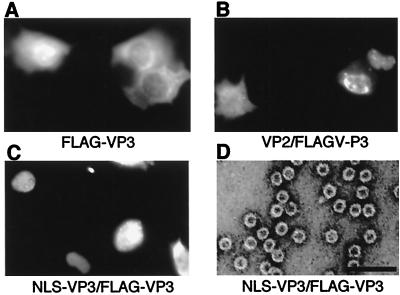
FIG. 4.
Nuclear transport and VLP formation of FLAG-VP3 in the presence of NLS-VP3. (A to C) Subcellular localization of FLAG-VP3 construct alone (A) or together with expression vectors containing VP2 (B) or NLS-VP3 (C). The localization of FLAG-VP3 was examined by indirect immunofluorescence staining. Cells were stained with anti-FLAG monoclonal antibody followed by rhodamine-conjugated mouse anti-immunoglobulin G. (D) Electron microscopy. Sf9 cells were infected with a recombinant baculovirus containing FLAG-VP3 and NLS-VP3. VLP purified by CsCl density gradient analysis was negatively stained and observed. Bar, 100 nm.

