Figure 1.
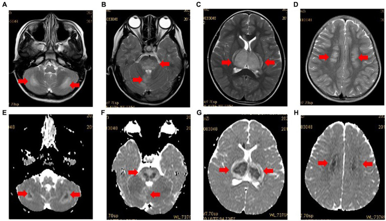
Case 3: MRI in case 3 with typical radiological findings of ANE. Axial T2-weighted images (A-D) showing bilateral symmetrical involvement of the cerebellum, brainstem, thalamus and cerebral white matter (arrows), respectively. The apparent diffusion coefficient (ADC) (E-H) presenting two gradient modes or there gradient modes.

