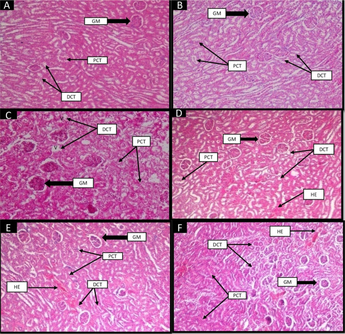
Figure 6.
Effect of camel milk and insulin on the histological structure of the kidney of albino Wistar rats (original magnification 10×). (A) Group I (control): H&E-stained section of the kidney showed the normal structure of glomerulus (GM), proximal convoluted tubules (PCT), and distal convoluted tubules (DCT). (B) Group II (control + camel milk): showed the normal structure of glomerulus, PCT, and DCT as in the control group. (C) Group III (diabetic control): STZ-induced diabetic group showed tubular (PCT and DCT) degeneration, vacuolization (V), and disruption of the glomerulus (GM). (D) Group IV (insulin): Reverted the damage of glomerulus (GM), improvisation in the structures of PCT and DCT. Vacuolization and tubular degeneration improved in this group. Slightly hemorrhage appeared in tubules. (E) Group V (camel milk): Depicted improvement in structures of the glomerulus, PCT, and DCT. Hemorrhage and slight vacuolization appeared in this group. (F) Group VI (camel milk + insulin): Showed slightly reverted structure of glomerulus, PCT, and DCT. Hemorrhages were present in tubules.

