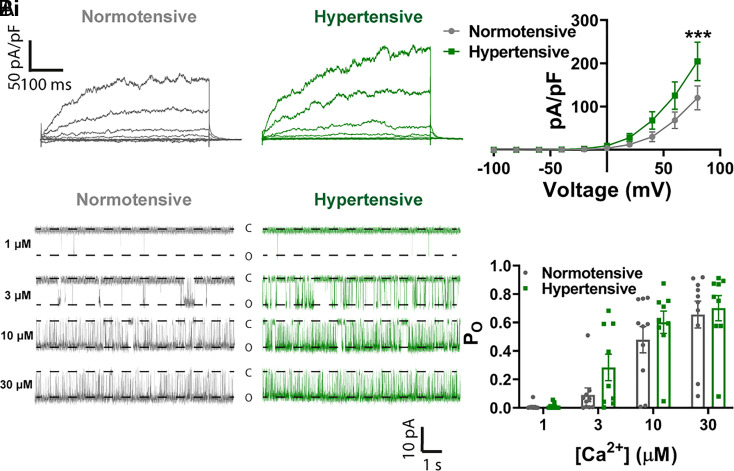
Fig. 4.
No decrease in functional BK subunit expression in hypertension. (A, i) Whole-cell ruptured-patch electrophysiological recordings of paxilline-sensitive currents obtained between −100 and 100 mV in SMCs isolated from pial arteries from normotensive (gray) and hypertensive (green) mice. (A, ii) Summary data comparing BK current density in cells isolated from hypertensive mice and normotensive mice (n = 14 arteries from 5 normotensive mice and 11 arteries from 4 hypertensive mice; *P < 0.05, **P < 0.01, two-way ANOVA). (B, i) Single-channel inside-out BK channel recordings at −40 mV in exercised patches from SMCs isolated from normotensive (gray) and hypertensive (green) pial arteries. Openings were evoked by exposing patches to 1 μM (Upper), 3 μM (Upper-Middle), 10 μM (Lower Middle), and 30 μM (Lower) free Ca2+. (B, ii) Summary data comparing the open probability (PO) between groups at different Ca2+ concentrations (n = 10 cells from 4 normotensive mice and 9 cells from 5 hypertensive mice; two-way ANOVA).

