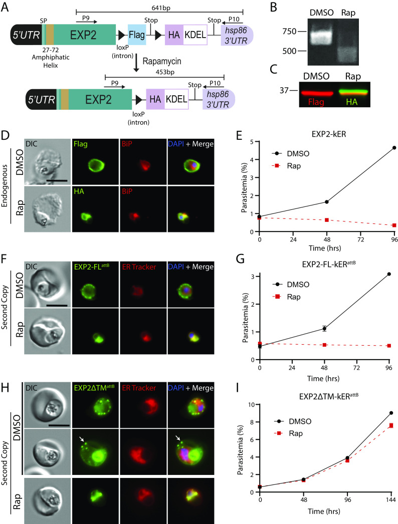
Fig. 3.
ER retention of EXP2 causes a lethal fitness defect independent of loss of PVM transport functions. (A) Schematic showing strategy for appending kER to the endogenous C-terminus of EXP2 without mNG. (B) PCR showing excision in EXP2-kER 24 h after treatment with rapamycin using primers P9/10. (C) Western blot of EXP2-kER parasites 24 h posttreatment with DMSO or rapamycin. Molecular weights after signal peptide cleavage are predicted to be 34.6 kDa for EXP2-3xFLAG and 34.7 kDa for EXP2-3xHA-KDEL. (D) IFA of EXP2-kER 24 h posttreatment with DMSO or rapamycin. (E) Growth of asynchronous parasites (n = 2 biological replicates) treated with DMSO or rapamycin. Data are presented as means ± SD from one biological replicate (n = 3 technical replicates). (F) Live microscopy of parasites expressing a full-length second copy of EXP2 with an mNG-kER fusion from the attB site under the control of the endogenous exp2 promoter (EXP2-FL-kERattB). Parasites were viewed 24 h after treatment with DMSO or rapamycin. (G) Representative growth of asynchronous EXP2-FL-kERattB parasites (n = 2 biological replicates) treated with DMSO or rapamycin. Data are presented as means ± SD from one biological replicate (n = 3 technical replicates). (H) Live microscopy of parasites expressing a second copy of EXP2 with mNG-kER fusion and lacking the amphipathic helix from the attB site (EXP2∆TM-kERattB). Parasites were viewed 24 h after treatment with DMSO or rapamycin. Two representative examples of unexcised parasites are presented, showing localization of the ∆TM version of EXP2 to the PV and host cell in infected RBCs (arrow). Digestive vacuole fluorescence was also observed as is typical for PV proteins. (I) Representative growth of asynchronous EXP2∆TM-kERattB parasites (n = 2 biological replicates) treated with DMSO or rapamycin. Data are presented as means ± SD from one biological replicate (n = 3 technical replicates). Second copy EXP2-kER lines were maintained in media supplemented with 500 nM aTc. (Scale bars, 5 μm.)

