Abstract
The endothelium of the normal corneas of 67 human subjects was studied in vivo with the specular microscope in order to quantify the method as a means of sampling the cell density of the tissue. It was found that (1) axial cell counts of the endothelium are reproducible in the same cornea after an interval of time; (2) the cell counts of the centre and periphery of the same cornea are similar; (3) the axial cell counts of pairs of eyes are similar; and (4) there is a gradual reduction of cell number with increasing age. The significance of these data is discussed.
Full text
PDF
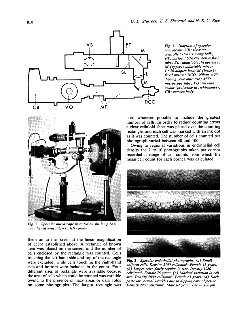
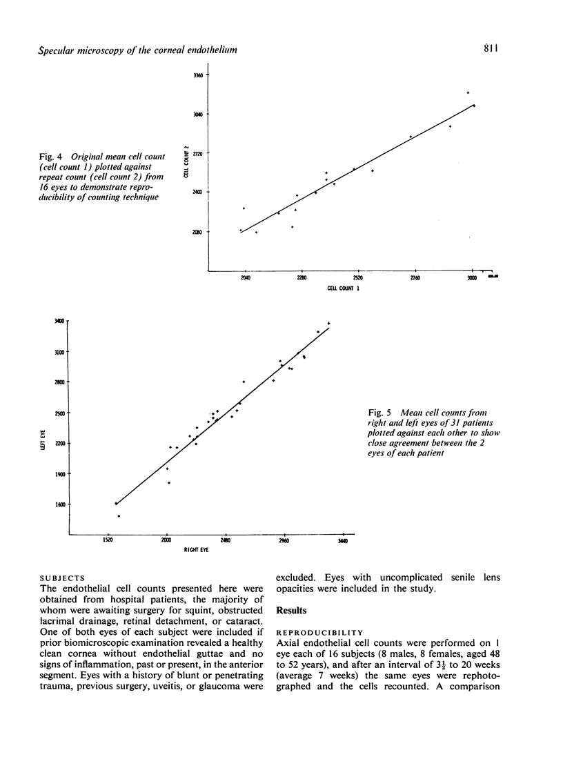
Images in this article
Selected References
These references are in PubMed. This may not be the complete list of references from this article.
- Binkhorst C. D., Loones L. H., Nygaard P. The clinical specular microscope. Doc Ophthalmol. 1977 Sep 30;44(1):57–75. doi: 10.1007/BF00171456. [DOI] [PubMed] [Google Scholar]
- Bourne W. M., Enoch J. M. Some optical principles of the clinical specular microscope. Invest Ophthalmol. 1976 Jan;15(1):29–32. [PubMed] [Google Scholar]
- Bourne W. M., Kaufman H. E. Cataract extraction and the corneal endothelium. Am J Ophthalmol. 1976 Jul;82(1):44–47. doi: 10.1016/0002-9394(76)90662-0. [DOI] [PubMed] [Google Scholar]
- Bourne W. M., Kaufman H. E. Endothelial damage associated with intraocular lenses. Am J Ophthalmol. 1976 Apr;81(4):482–485. doi: 10.1016/0002-9394(76)90305-6. [DOI] [PubMed] [Google Scholar]
- Bourne W. M., Kaufman H. E. Specular microscopy of human corneal endothelium in vivo. Am J Ophthalmol. 1976 Mar;81(3):319–323. doi: 10.1016/0002-9394(76)90247-6. [DOI] [PubMed] [Google Scholar]
- Bourne W. M., McCarey B. E., Kaufman H. E. Clinical specular microscopy. Trans Sect Ophthalmol Am Acad Ophthalmol Otolaryngol. 1976 Sep-Oct;81(5):743–753. [PubMed] [Google Scholar]
- IRVINE A. R., IRVINE A. R., Jr Variations in normal human corneal endothelium; a preliminary report of pathologic human corneal endothelium. Am J Ophthalmol. 1953 Sep;36(9):1279–1285. doi: 10.1016/0002-9394(53)92298-3. [DOI] [PubMed] [Google Scholar]
- IRVINE A. R., Jr The role of the endothelium in bullous keratopathy. AMA Arch Ophthalmol. 1956 Sep;56(3):338–351. doi: 10.1001/archopht.1956.00930040346003. [DOI] [PubMed] [Google Scholar]
- Kaufman H. E., Capella J. A., Robbins J. E. The human corneal endothelium. Am J Ophthalmol. 1966 May;61(5 Pt 1):835–841. doi: 10.1016/0002-9394(66)90921-4. [DOI] [PubMed] [Google Scholar]
- Laing R. A., Sandstrom M. M., Leibowitz H. M. In vivo photomicrography of the corneal endothelium. Arch Ophthalmol. 1975 Feb;93(2):143–145. doi: 10.1001/archopht.1975.01010020149013. [DOI] [PubMed] [Google Scholar]
- Laing R. A., Sandstrom M., Berrospi A. R., Leibowitz H. M. Morphological changes in corneal endothelial cells after penetrating keratoplasty. Am J Ophthalmol. 1976 Sep;82(3):459–464. doi: 10.1016/0002-9394(76)90495-5. [DOI] [PubMed] [Google Scholar]
- Laing R. A., Sanstrom M. M., Berrospi A. R., Leibowitz H. M. Changes in the corneal endothelium as a function of age. Exp Eye Res. 1976 Jun;22(6):587–594. doi: 10.1016/0014-4835(76)90003-8. [DOI] [PubMed] [Google Scholar]
- Maurice D. M. Cellular membrane activity in the corneal endothelium of the intact eye. Experientia. 1968 Nov 15;24(11):1094–1095. doi: 10.1007/BF02147776. [DOI] [PubMed] [Google Scholar]
- Sherrard E. S. The corneal endothelium in vivo: its response to mild trauma. Exp Eye Res. 1976 Apr;22(4):347–357. doi: 10.1016/0014-4835(76)90227-x. [DOI] [PubMed] [Google Scholar]
- Svedbergh B., Bill A. Scanning electron microscopic studies of the corneal endothelium in man and monkeys. Acta Ophthalmol (Copenh) 1972;50(3):321–336. doi: 10.1111/j.1755-3768.1972.tb05955.x. [DOI] [PubMed] [Google Scholar]


